Upper Leg Tendon Anatomy ~ Two Jointed Muscles Of The Lower Body What They Are And How To Train Breaking Muscle. Lie prone on a hamstring curl machine. Learn vocabulary, terms and more with flashcards, games and other study tools. Posterior surface of calcaneus (via calcaneal tendon). Use the mouse scroll wheel to move the images up and down alternatively use the tiny arrows (>>) on both side of the image to move the images. Find stockbilleder af concept 3d human upper leg anatomy i hd og millionvis af andre royaltyfri stockbilleder, illustrationer og vektorer i shutterstocks samling.
The pads of the machine are situated at the achilles tendon. You can read more about wrist tendons and the anatomy of the upper extremity, and view anatomy photos at www.handcare.org. Try to do both of these exercise on your own, before i post my answer briefly, it sits inside the quadriceps tendon and connects it to the front of the tibia by way of the patellar ligament. The thigh bone, or femur, is the large upper leg bone that connects the lower leg bones (knee joint) to the pelvic bone (hip joint). Hands are outstretched, holding onto the handles of the bench.
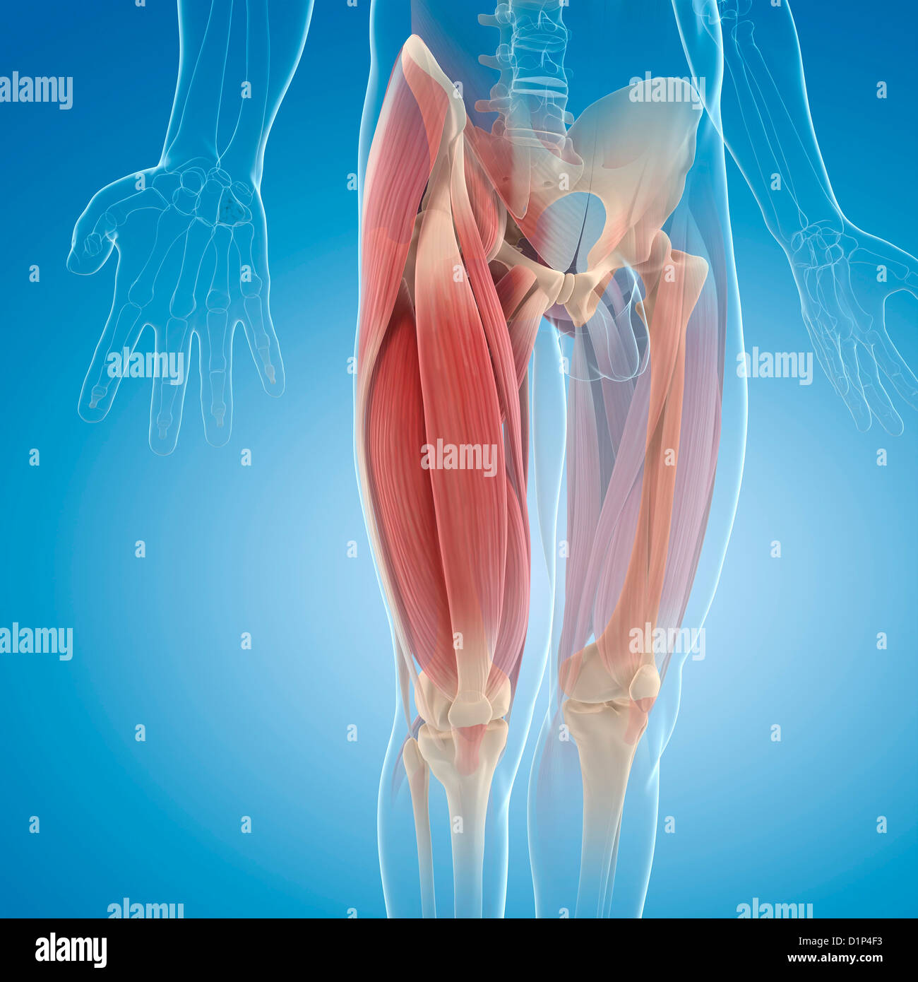
Leg Anatomy All About The Leg Muscles from www.kingofthegym.com Do anatomy tracings over those to find the leg bones. Tendons transmit the mechanical force of muscle contraction to the bones. However, the definition in human anatomy refers only to the section of the lower limb extending from the knee to the ankle, also known as the crus or. For more on tendon anatomy, refer here. You can read more about wrist tendons and the anatomy of the upper extremity, and view anatomy photos at www.handcare.org. There is no real division between the core and the upper leg; The lower leg is comprised of two bones, the tibia and the smaller fibula. Fibula— a long, thin bone in the lower leg on the lateral side which runs along side the tibia from the knee to the ankle.
The lower leg is comprised of two bones, the tibia and the smaller fibula.
This is the physiological upper limit of tendon strain whereby the collagen fibrils orient themselves in the direction of tensile mechanical. This mri wrist coronal cross sectional anatomy tool is absolutely free to use. The anatomical basis of clinical practice. Use the mouse scroll wheel to move the images up and down alternatively use the tiny arrows (>>) on both side of the image to move the images. The human leg, in the general word sense, is the entire lower limb of the human body, including the foot, thigh and even the hip or gluteal region. The tendons that control movement in your hands, wrists and fingers run through your forearm. Enters the anterior compartment of the leg through a gap in the upper part of the interosseous membrane. To describe the mechanical properties of tendons. See the pictures and anatomy description of knee joint bones, cartilage, ligaments, muscle and tendons with resources for knee problems & injuries. Want to learn more about it? Fibula— a long, thin bone in the lower leg on the lateral side which runs along side the tibia from the knee to the ankle. Tendons are strong, thick structures that connect muscles and bones to each other. These nerves give sensation to our upper.
The peroneal tendons are in the feet and provide balance and stability during movement. Tendons are also bands of connective tissue. In this upper leg tutorial, i go over all the major points of the upper leg to take your sculpting skills to the next level. Extends leg at knee in quad group. Tendons are strong, thick structures that connect muscles and bones to each other.
Natural Color Photograph Of Dissection Of The Left Upper Leg Posterior View Showing The Exiting Of The Sciatic Nerve Below The Piriformis Muscle And The Related Structures Orthopaedic Surgical Anatomy Teaching from digitallibrary.usc.edu The tendons that control movement in your hands, wrists and fingers run through your forearm. Your biceps tendons attach the biceps muscle to bones in your shoulder and in your elbow. Tendons are also bands of connective tissue. There are 7 main areas covered in the upper limb; However, the definition in human anatomy refers only to the section of the lower limb extending from the knee to the ankle, also known as the crus or. Find stockbilleder af concept 3d human upper leg anatomy i hd og millionvis af andre royaltyfri stockbilleder, illustrationer og vektorer i shutterstocks samling. Try to do both of these exercise on your own, before i post my answer briefly, it sits inside the quadriceps tendon and connects it to the front of the tibia by way of the patellar ligament. Try this movement out by standing on one foot with the other leg.
Tendons are fibrous cords attached to muscles and bone.
Tendons are fibrous cords attached to muscles and bone. Tendons are fibrous cords, similar to a rope, and are made of collagen. The image is available for download in high resolution quality up to 2946x2946. This mri wrist coronal cross sectional anatomy tool is absolutely free to use. There are 7 main areas covered in the upper limb; It's the area that runs from the hip to the knee in each leg. Do anatomy tracings over those to find the leg bones. The muscles, bones, joints, nerves, blood and lymphatic supply, anatomical areas, and the the nerves of the upper limb arise from a complex arrangement of nerve fibers known as the brachial plexus; Look for subcutaneous landmarks to figure out where the bones go. Hands are outstretched, holding onto the handles of the bench. See the pictures and anatomy description of knee joint bones, cartilage, ligaments, muscle and tendons with resources for knee problems & injuries. This is the physiological upper limit of tendon strain whereby the collagen fibrils orient themselves in the direction of tensile mechanical. Iliotibial band syndrome description the iliotibial band is the tendon attachment of hip muscles into the upper leg (tibia) just below the knee to the outer side of the front of the leg.
The image is available for download in high resolution quality up to 2946x2946. This is the physiological upper limit of tendon strain whereby the collagen fibrils orient themselves in the direction of tensile mechanical. This mri wrist coronal cross sectional anatomy tool is absolutely free to use. Peroneal tendonitis affects these tendons, and can make movement difficult and painful. There are 7 main areas covered in the upper limb;
Human Upper Leg Muscles High Resolution Stock Photography And Images Alamy from c8.alamy.com Medically reviewed by william morrison, m.d. Look for subcutaneous landmarks to figure out where the bones go. It's the area that runs from the hip to the knee in each leg. Try to do both of these exercise on your own, before i post my answer briefly, it sits inside the quadriceps tendon and connects it to the front of the tibia by way of the patellar ligament. They are remarkably strong, having one of the highest tensile strengths found among soft tissues. There are 7 main areas covered in the upper limb; Want to learn more about it? There is no real division between the core and the upper leg;
Learn about the causes, treatments, and outlook for this condition.
The image is available for download in high resolution quality up to 2946x2946. Enters the anterior compartment of the leg through a gap in the upper part of the interosseous membrane. This mri wrist coronal cross sectional anatomy tool is absolutely free to use. Extends leg at knee in quad group. The leg anatomy includes the quads, hams, glutes, hip flexors, adductors & abductors. Posterior surface of calcaneus (via calcaneal tendon). The anatomical basis of clinical practice. The tendons that control movement in your hands, wrists and fingers run through your forearm. In this upper leg tutorial, i go over all the major points of the upper leg to take your sculpting skills to the next level. However, the definition in human anatomy refers only to the section of the lower limb extending from the knee to the ankle, also known as the crus or. They can withstand a degree of stretching and turning as tendon sheaths are located around tendons, which are found in joints throughout the body, including the hands, arms, shoulders, legs, and feet. Yoga anatomy anatomy study anatomy reference. Quadriceps tendon to base of patella and onto tibial tuberosity via the patellar ligament action:
Share :
Post a Comment
for "Upper Leg Tendon Anatomy ~ Two Jointed Muscles Of The Lower Body What They Are And How To Train Breaking Muscle"


Post a Comment for "Upper Leg Tendon Anatomy ~ Two Jointed Muscles Of The Lower Body What They Are And How To Train Breaking Muscle"